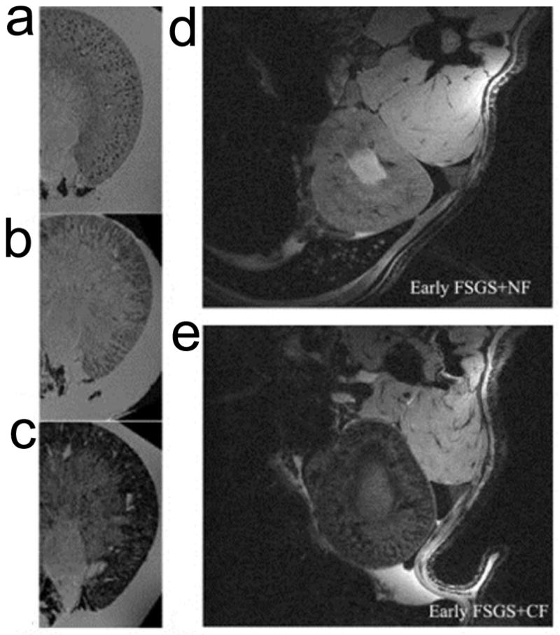
Figure 2. Detection of glomerular injury in puromycin-induced focal and segmental glomerulosclerosis (FSGS) by MRI with cationized ferritins (CF).

a, Ex vivo MRI on normal kidneys, CF was injected beforehand. Spotted distribution of glomeruli was observed. b, Ex vivo MRI on kidneys from early FSGS, which was induced by injection of puromycin amnionucleoside (PAN). The spots were still visible but were surrounded by areas of signal hypointensity. c, The spots were no longer visible for late FSGS due to enlarged areas of hypointensities. d, In vivo MRI of early FSGS after injection of native ferritins (NF). e, In vivo MRI of early FSGS after injection of CF. Reprinted with permission from ref [39].
