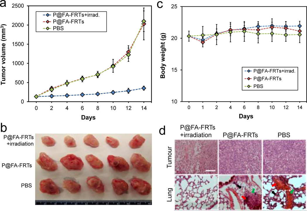
Fig. 4.
(a) Tumour growth curves. Significant tumour suppression was observed in animals treated with P@FA-FRT-mediated PDT. Compared to the control group, a tumour growth suppression rate of 82.65 ± 4.11 % was observed on day 14. (b) Photographs of dissected tumours from (a). (c) Body weight curves. No significant weight loss was observed for the treatment group. (d) H&E staining with tumour (upper) and lung (lower) samples. Significant necrosis was observed in tumours treated with P@FA-FRT-mediated PDT. In addition, while the control groups showed signs of metastasis in the lung (e.g. thickened alveolar membranes [black arrows], bleeding [red arrows], and inflammation sites [green arrows]), there was no sign of lung metastasis for PDT treated animals. Scale bar, 100 µm.

