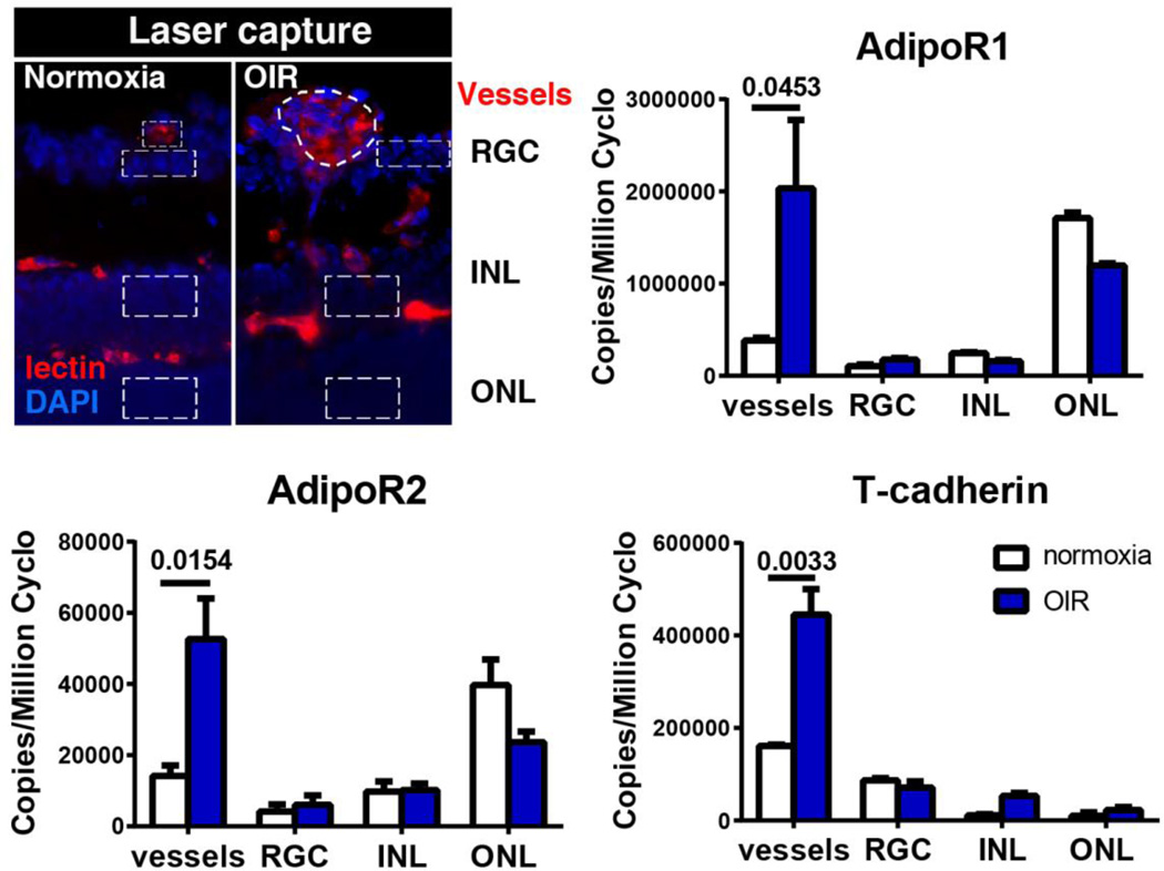
Figure 2.
Expression of APN receptors in retinal neuronal layers and blood vessels in normal and hypoxic retinas at P17, when maximal neovascularization is observed in the mouse model of oxygen-induced retinopathy. Schematic of the laser-capture microdissected retinal layers (DAPI for nuclei, blue) and blood vessels (lectin, red) is shown (outlined with dotted line). Expression of adipoR1, adipoR2, and T-cadherin was examined by using qPCR. GCL, ganglion cell layer; INL, inner nuclear layer; ONL, outer nuclear layer.
Adapted from Fu et al, AJCN, 2015, 101 (4): 879–88

