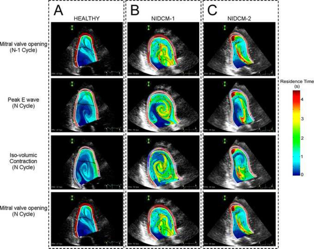
FIGURE 2.
Snapshots of 2-D intraventricular residence time along the cardiac cycle in a healthy heart (A) and in two different examples of dilated cardiomyopathy (NIDCM) patients (B & C). 1st row: Residence Time mapping at the mitral valve opening in the converged N-l cycle. 2nd row: Residence Time mapping at peak E-wave in the last computed cycle. 3rd row: Residence Time mapping at the iso-volumetric contraction in the last computed cycle. 4th row: Residence Time mapping at mitral valve opening in the last cycle. Notice that in both NIDCMs there coexist different regions with high Tr.

