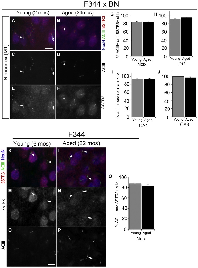
Figure 2.
Somatostatin receptor 3 (SSTR3)-positive cilia in young and aged rat forebrain. (A–F) Epifluorescent images of 2 and 34 mos F344 × BN layer 2/3 primary motor neocortex (M1) co-immunolabeled for ACIII (green), SSTR3 (red) and NeuN (blue). Arrowheads indicate cilia positive for both SSTR3 and ACIII. (G–J) Percent of NeuN+ neurons that possess SSTR3+ cilia in M1 (G) and hippocampal DG (H), CA1 (I) and CA3 (J) of 2 and 34 mos F344 × BN rats. (K–P) Images of 6 (left panels) and 24 mos (right panels) F344 rat layer 2/3 frontal neocortex immunostained for SSTR3 (red), ACIII (green) and NeuN (red). The arrowheads indicate cilia positive for both SSTR3 and ACIII, while the arrows (A,C,E,L,N,P) show examples of an ACIII positive cilium that is SSTR3 negative. (Q) Percent of NeuN positive neurons that possess SSTR3 positive cilia in frontal neocortex. Scale bars (A,O) = 10 μm.

