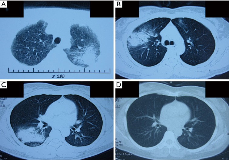
Figure 2.
Chest computed tomography (CT) scan presenting as pulmonary migratory infiltrates (PMI). (A) CT scan after the first hospitalization indicates a new lesion of consolidation in the left upper lobe; (B) CT scan on the third presentation shows patchy opacification with air bronchogram in the right upper lung field and small area of ground-glass opacity in the left upper field; (C) CT scan after the third hospitalization shows patchy opacification in the right lower lobe; (D) CT scan obtained after 7 days treatment of azithromycin shows complete resolution of the lesion in the right lower lobe.

