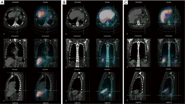
Figure 3.
Fused SPECT-CT imaging. (A) 99mTc-nanocolloid scan shows uptake in paracardiac solid mass, confirming the diagnosis of thoracic splenosis (TS) (left panel CT scan, right panel fused images); (B) 99mTc-nanocolloid scintigraphy (left panel CT scan, right panel fused images) reveals radiotracers uptake in the pleural nodule of the left hemithorax; (C) the solid mass in the splenic space shows radiotracer uptake at 99mTc-nanocolloid scintigraphy (left panel CT scan, right panel fused images). SPECT, single photon emission computed tomography; CT, computed tomography.

