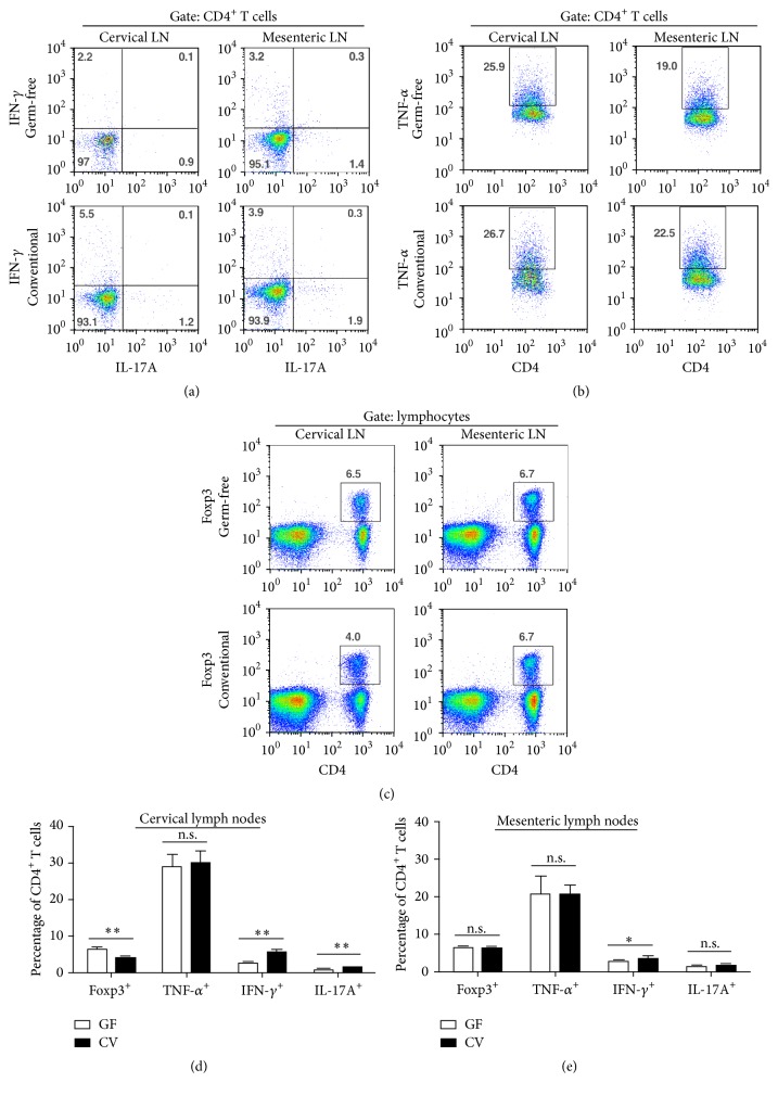
Figure 5.
Flow cytometric analysis of lymphocyte populations in cervical and mesenteric lymph nodes. In the cervical lymph nodes of conventional (CV) mice, (a) the percentage of IFN-γ and IL-17-producing CD4+ T cells increased and (c) the percentage of regulatory Foxp3-expressing CD4+ T cells decreased compared to germ-free (GF) mice. In the mesenteric lymph nodes of CV mice, the environment was less proinflammatory showing only a small but significant increase of IFN-g-producing CD4+ T cells compared to GF mice. In both cervical and mesenteric lymph nodes, (b) the percentage of TNF-α-producing CD4+ T cells remained unchanged. The dot plots are representative of two independent experiments. The column graphs summarize the frequency of lymphocyte subpopulations in (d) the cervical and (e) the mesenteric lymph nodes. Each graph represents data from two independent experiments. ∗ p < 0.05, ∗∗ p < 0.01 (Mann-Whitney test). n.s. = not significant.

