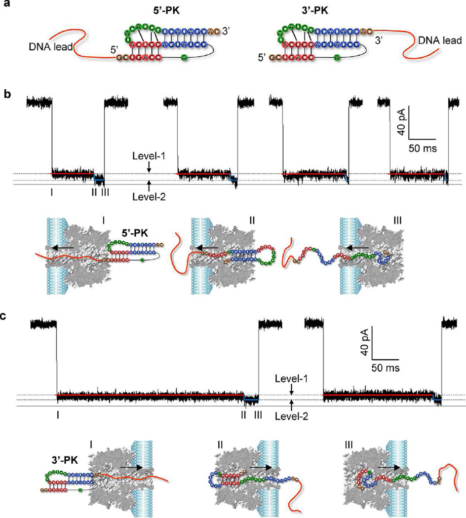
Figure 2.
Nanopore signatures revealing vectorial unfolding procedure of T2 pseudoknot. (a) Construction of DNA-tagged pseudoknot chimeras. A poly(CAT)10 ssDNA is attached to the 5′ end (5′-PK) and 3′ end (3′-PK) of T2 pseudoknot for trapping the molecule into the pore and drive its unfolding. (b,c) Representative two-level signatures (Level-1 and Level-2) for stepwise unfolding of 5′-PK from H1 at 5′ end to H2 at 3′ end (b), and 3′-PK from H2 at 3′ end to H1 at 5′ end (c). Models below the current traces illustrate the suggested molecular configurations of 5′-PK (b) and 3′-PK (c) for each unfolding step. The nanopore currents were recorded at +120 mV in 1 M NaCl and 10 mM MgCl2 buffered with 25 mM MOPS (pH 7.4).

