Figure 4.
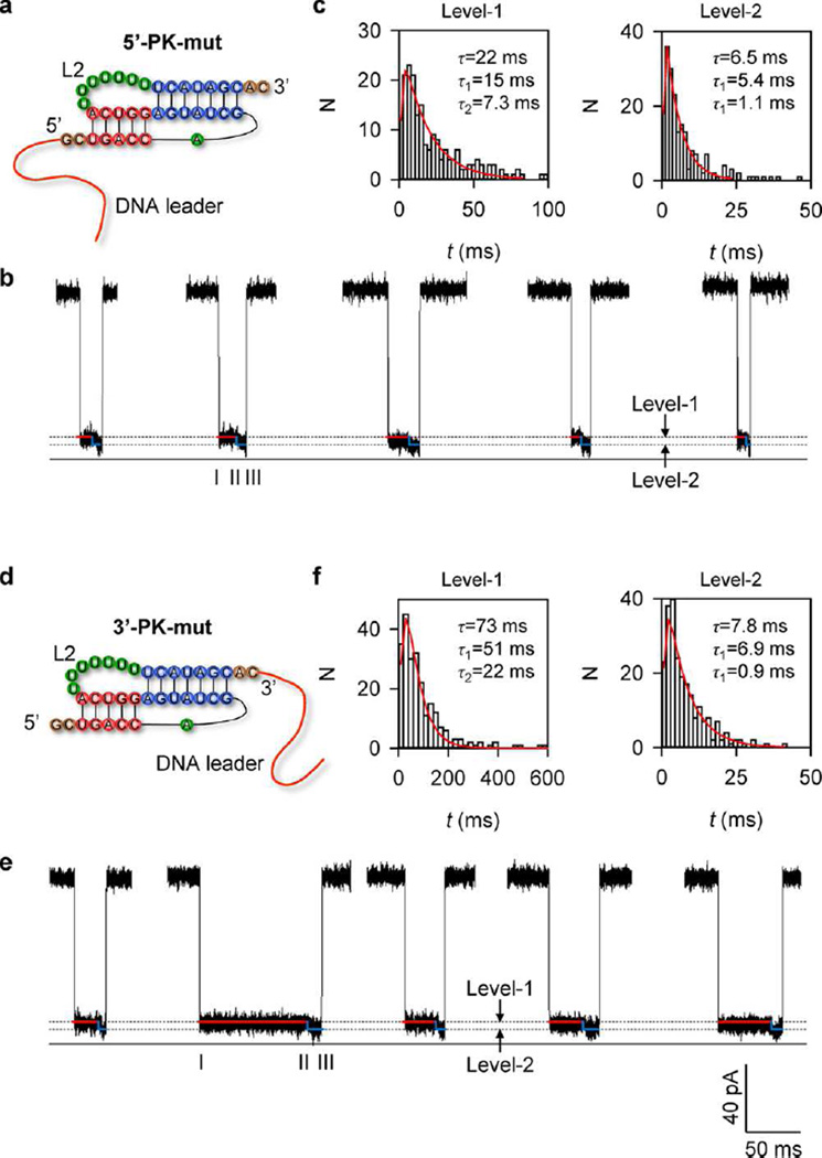
Unfolding of 5′- and 3′-PK-mut in the nanopore. (a) 2D structure of 5′-PK-mut, with L2 changed to poly(U). (b) Representative two-level signatures (Level-1 and Level-2) for stepwise unfolding of 5′-PK-mut from H1 at 5′ end to H2 at 3′ end. (c) Representative histograms of Level-1 (left) and level-2 (right) durations (N = 252), which were both fitted with a two-step exponential distribution as 5′-PK in Figure 3. (d) 2D structure of 3′-PK-mut. (e) Representative two-level signatures (Level-1 and Level-2) for stepwise unfolding of 3′-PK-mut from H2 at 3′ end to H1 at 5′ end. (f) Representative histograms of Level-1 (left) and Level-2 (right) durations (N = 239) and the two-step exponential fitting as 3′-PK in Figure 3.

