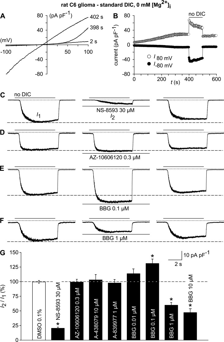
Figure 10.
Electrophysiological TRPM7 fingerprint and effects of P2X7 receptor antagonist on TRPM7-like currents in rat C6 glioma cells. (A) Whole-cell currents were measured during repetitive (every 2 s) application of voltage ramps (−100 to 100 mV; 0.8 mV/ms) under standard DIC conditions at 2 and 398 s after the start of the experiment and after switching to a no-DIC bath solution (402 s). (B) Statistical evaluation of seven experiments performed as in A. Shown are inward current amplitudes at −80 mV (filled circles) and outward current amplitudes at 80 mV (open circles) versus time. (C–F) TRPM7-like whole-cell currents at −60 mV, recorded after preconditioning (not depicted), were elicited by repetitive exposure (6 s at 30-s intervals) to no DIC before (left; I1) and 6 min after exposure to the indicated TRPM7 and P2X modulators (middle; I2). (right) Currents 6 min after wash-out of the test compounds. (G) Statistical analysis of experiments as in A–E (n = 7–9 each). TRPM7-like peak currents in the presence of the test compounds (I2) are normalized to the respective pre-application peak currents (I1). *, P < 0.001, significantly different from the control condition (DMSO). In electrophysiological figures, dotted lines indicate the zero current level. Error bars indicate SEM.

