Figure 3.
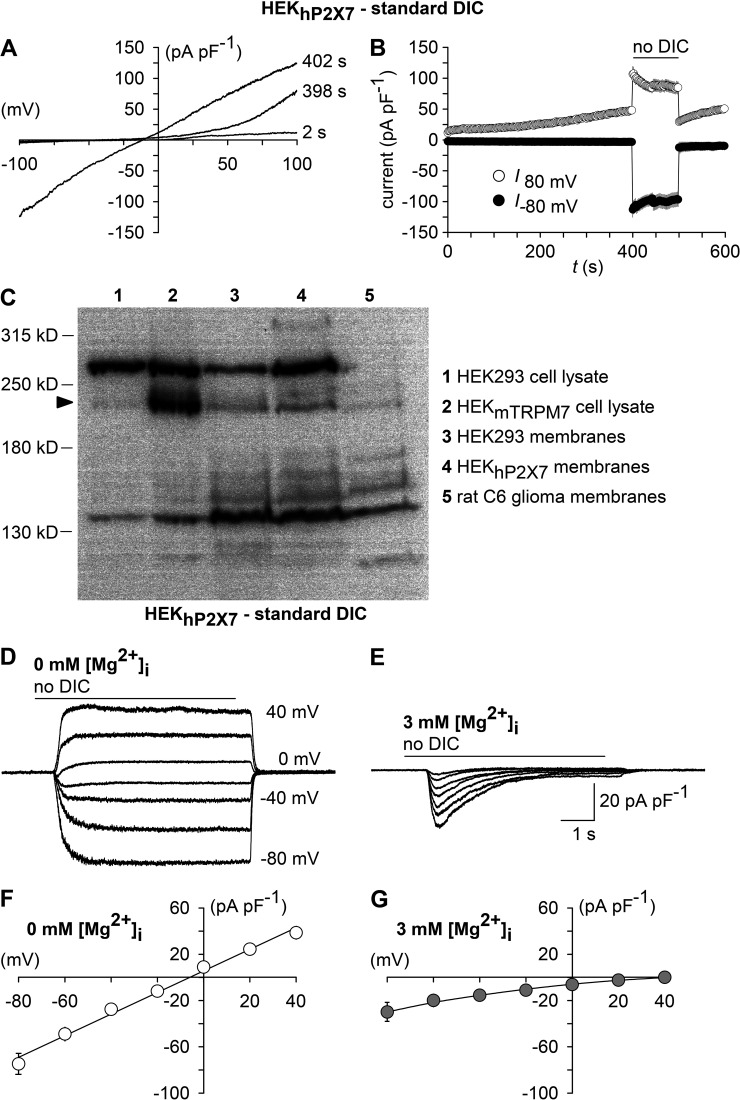
TRPM7 fingerprinting. (A) Shown are superimposed currents in a HEKhP2X7 cell during application of voltage ramps (0.8 mV/ms) under standard DIC conditions at 2 and 398 s after the start of the experiment and, additionally, at 402 s, when the extracellular medium had been exchanged to no DIC. The holding potential between ramps was 0 mV. (B) Statistical evaluation of seven experiments performed as shown in A. Depicted are inward current amplitudes at −80 mV (filled circles) and outward current amplitudes at 80 mV (open circles) versus time. Note the run-up of outward currents, as well as the augmentation of inward currents associated with a linearization of the previously outwardly rectifying I/V curve in the absence of extracellular DICs (no DIC), which are characteristic features of TRPM7. (C) Western blot analysis of TRPM7 expression in membrane fractions of parental HEK293 cells (3), HEKhP2X7 cells (4), and rat C6 glioma cells (5). Blots were probed with recombinant monoclonal anti-TRPM7.Whole-cell lysates of HEK293 cells transiently transfected with an expression plasmid encoding mouse TRPM7 (HEKmTRPM7, 2) were used to visualize the position of the TRPM7-specific band (arrowhead), and results from the respective untransfected HEK293 clone 1 are shown for comparison. (D and E) Superimposed whole-cell currents from HEKhP2X7 cells recorded with a Mg2+-free (D) or 3 mM Mg2+–containing pipette solution (E), showing the effect of no-DIC bath, which was repeatedly applied 6 s at 30-s intervals at varying (20-mV increments) membrane voltages. Note that sustained responses were abolished by 3 mM [Mg2+]i. (F and G) Current-voltage curves constructed from experiments similar to those in D and E (n = 11 each). Error bars indicate SEM.

