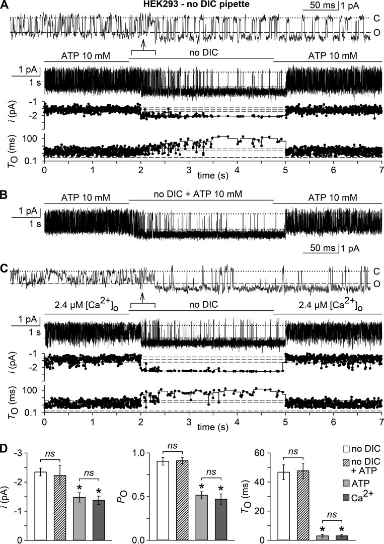
Figure 6.
Extracellular Ca2+ but not ATP apparently blocks TRPM7. (A–C) Shown are currents in outside-out membrane patches excised with a pipette containing no-DIC solution from HEK293 cells. Currents were evoked either by low DIC plus 10 mM ATP (2.4 µM free [Ca2+]o; A and Β) or by a bath solution containing only 2.4 µM free [Ca2+]o (C). Channel activity was then modulated by switching to no DIC, without (A and C) or with added ATP (10 mM; B). Top panels in A and C show unitary current activity at a higher time resolution from the data sections specified by arrows. Bottom panels in A and C illustrate stability plots of unitary current amplitudes (i) and open times (TO), when plotted versus time on the same abscissa scaling as the respective current traces. Broken lines indicate the mean and standard deviation of i or TO during the 1.75 s of recording before the bath medium was exchanged. (D) Means ± SEM of unitary current amplitudes (i), intraburst open probabilities (PO), mean intraburst closed times (TC), and mean intraburst open times (TO) of the channels activated by no DIC (open bars), no DIC plus 10 mM ATP (hatched bars), ATP (in low DIC; 2.4 µM free [Ca2+]o; light gray bars), and 2.4 µM [Ca2+]o (dark gray bars). *, P < 0.001, significantly different from the channel activity evoked by no DIC; ns, not statistically significant; n = 7–9 each. Single channel parameters were derived from a 1.75-s recording period before bath exchange (ATP in low DIC, 2.4 µM [Ca2+]o) or from the last 2.5 s in no DIC and ATP in no DIC, respectively, and included 65–983 dwells. In electrophysiological figures, dotted lines indicate the zero current level.

