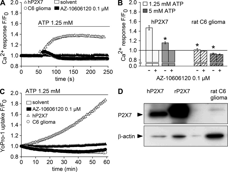
Figure 8.
Lack of functional P2X7 expression in rat C6 glioma cells. HEKhP2X7 (triangles) and rat C6 glioma cells (circles) were subjected to Ca2+ and Yo-Pro-1 uptake assays and probed for P2X7 expression. (A) Fluo-4/AM–loaded cells were incubated with 0.1 µM AZ10606120 (filled symbols) or solvent (open symbols) for 5 min, and Fluo-4 fluorescence was detected before and after injection of 1.25 mM ATP as indicated. Fluorescence intensities (F) were normalized to the respective initial intensities (F0), and representative examples for HEKhP2X7 and rat C6 glioma cells are shown. (B) Statistical analysis (means ± SEM) of four to five independent measurements performed as shown in A or with a higher ATP concentration (5 mM, gray bars). Asterisks indicate significant differences (*, P < 0.05) compared with HEKhP2X7 cells stimulated with 1.25 mM ATP. (C) Yo-Pro-1 fluorescence was measured in HEKhP2X7 and in C6 glioma cells in low-DIC buffer, containing 0.1 mM CaCl2 and no MgCl2, supplemented with 0.1 µM AZ10606120 or solvent 5 min before stimulation with 1.25 mM ATP. Shown are background-corrected fluorescence intensities normalized to the initial intensity (F0). (D) Immunoblot analysis of P2X7 and β-actin in whole-cell lysates from HEK293 cells stably expressing human (hP2X7) or rat (rP2X7) P2X7 and from rat C6 glioma cells. Arrowheads indicate the expected molecular mass of human P2X7 (69 kD) and β-actin (42 kD).

