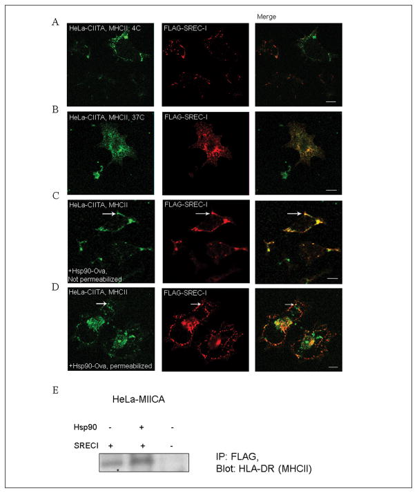
Figure 5. SREC-I co-localizes with MHCIIA on the cell surface and in intracellular organelles.
A, B) HeLa cells stably expressing Myc-CIITA were transfected with FLAG-SREC-I for 18 hours and then incubated at 4°C (A), or 37°C (B). Cells were then fixed and stained for SREC-I (FLAG-SREC-I, red) and MHCII ab (MHCII, green) (A) or permeabilized and stained as in (A). C, D) HeLa-CIITA cells were again transfected as in 5A and then incubated with Hsp90-Ova for 5 minutes. Cells were then stained for MHCII (MHCII, green) and FLAG (red) without permeabilization for cell surface localization assay. D) Transfected HeLa cells (as in A, B) were incubated with Hsp90-Ova for 15 minutes on ice and then incubated with warm growth media at 37°C. Cells were permeabilized and stained for MHCII (green) and FLAG (red). E) HeLa-FLAG-SREC-I and HeLa cells were transfected with CIITA-myc for 22 hours. Transfected and non-transfected HeLa cells were incubated with or without Hsp90.Ova (Hsp90) for 30 minutes. Cells were then incubated with anti-FLAG antibody beads for overnight at 4°C. Cell precipitates were collected, lysed and run for SDS-PAGE and later blotted for MHCII with anti-HLA-DR ab. Scale bar 5μm.

