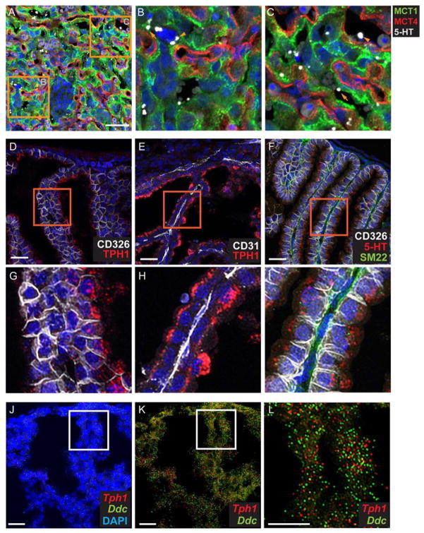
Fig 3. TPH1 and 5-HT are present in the visceral endoderm yolk sac.
(A–C) 5-HT is present in all cell types in the labyrinth, as well as in the fetal blood space. Images of CoIF staining with anti-5HT, anti-MCT1, anti-MCT4 performed on E12.5 placenta. (B and C), higher magnification images of the boxed area in (A). In (B), arrowhead points to a S-TGC apposed to SynT-I. In (C), arrowhead points to fetal blood space and arrow points to MCT1+ SynT-I. Scale bar in A = 50 μm. (D–I) Co-IF staining was performed with anti-5HT and yolk sac cell type specific molecular markers, CD326, visceral endoderm marker, CD31, endothelial cell marker, and SM22, mesoderm marker. (G–I), higher magnification of boxed area of (D–H) respectively. Scale bar in D to F = 30 μm. (J–L) Multiplex fluorescent ISH using RNAscope probes directed against Tph1 and Ddc performed on the same section. (L), higher magnification of boxed area in J and K. Blue: DAPI. Scale bar = 30 μm.

