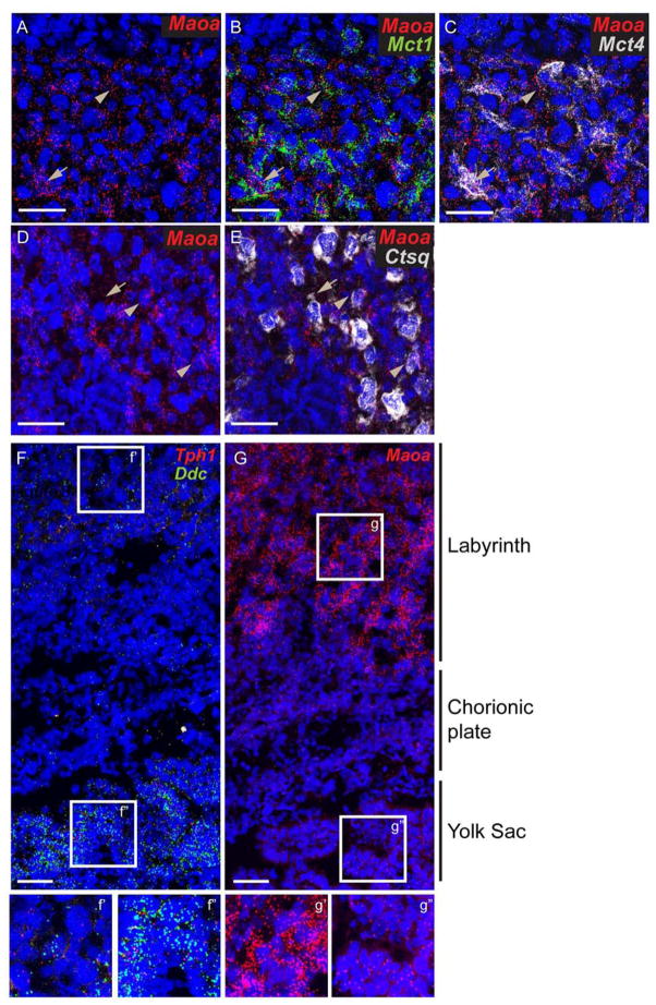
Fig 5. Expression of Maoa in the labyrinth and yolk sac.
Multiplex fluorescent ISH using RNAscope probes directly against (A–C) Maoa, Mct1, and Mct4 on the same section, (D–E) Maoa and Ctsq on the same section, (F–G) Maoa, or Ddc, and Tph1 (on the same section) on E14.5 placenta sections. (f’, f’’, g’, and g’’) magnify view of boxed region in F and G. Blue: DAPI. Scale bar = 50 μm.

