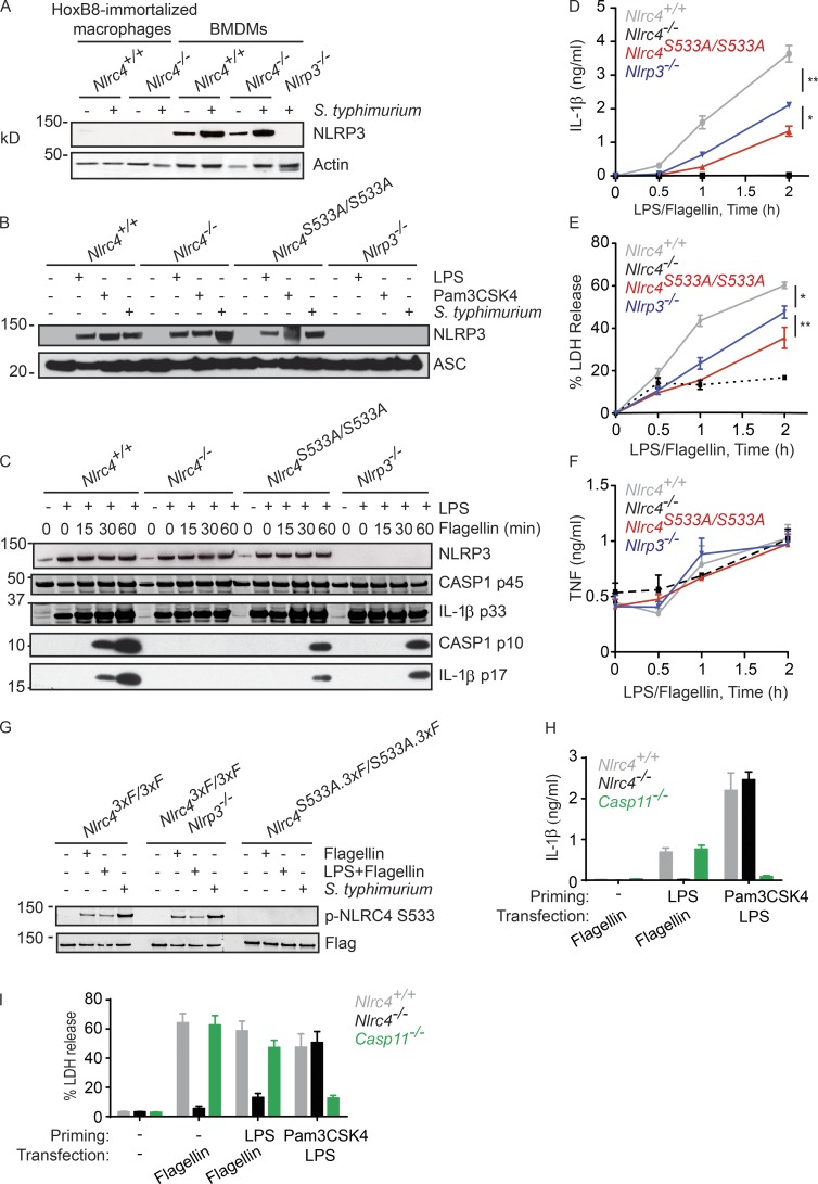
Figure 2.
NLRP3, but not caspase-11, contributes to NLRC4-dependent activation of caspase-1. (A and B) Western blots of immortalized macrophages (A) or BMDMs (A, B). Cells were primed for 4 h with LPS or Pam3CSK4, or infected for 2.5 h with S. typhimurium (MOI = 5). (C) Western blots of BMDMs and their culture medium. (D–F) Graphs show IL-1β (D), LDH (E) and TNF (F) released from BMDMs. (G) Western blots of NLRC4 immunoprecipitated with anti-FLAG antibody from BMDMs at 4 h after either infection (MOI = 10) or flagellin transfection. p-NLRC4, phosphorylated NLRC4. (H and I) Graphs show IL-1β (H) and LDH (I) released from BMDMs. Graphed data (mean ± SD) are pooled from three independent experiments for a total of three mice of each genotype. P-values were determined by the Student’s t test. *, P < 0.05; **, P < 0.01.

