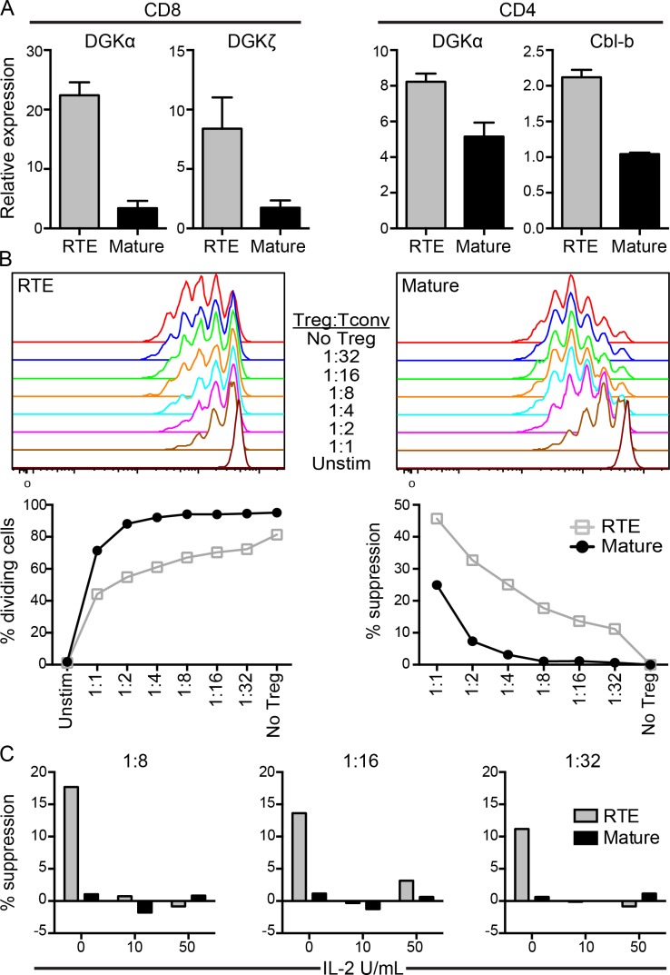
Figure 3.
RTEs express elevated levels of anergy-associated genes after in vivo encounter with self-antigen and are more susceptible to T reg cell–mediated suppression. (A, left two graphs) 106 naive OT-I RTEs and MN T cells were cotransferred into RIP-mOVA Tg hosts. (A, right two graphs) 1–2 × 10⁶ naive OT-II RTEs and MN T cells were cotransferred with 2 × 10⁶ bulk naive OT-I T cells into RIP-mOVA Tg hosts. Antigen-exposed (CD44hi) donor OT-II and OT-I T cells were sorted 7 d later from pLNs and RNA was extracted for gene expression analyses. Data are presented as mean ± SD and are representative of two independent experiments. (B and C) 105 naive polyclonal CFSE-labeled CD4 RTEs or MN T cells were co-cultured in vitro with the indicated ratios of Foxp3+ T reg cells in the presence of 105 irradiated TCRβ/δ−/− splenocytes without (B) or with (C) recombinant IL-2. Cells were cultured for 72 h without (unstim) or with 50 ng/ml soluble anti-CD3 and CFSE dilution measured (top). (B) Data in the bottom left panel show the percentage of dividing cells and in the bottom right panel show the percentage of suppression of T cell proliferation by Foxp3+ T reg cells. Data are representative of three independent experiments. (C) Data show the percentage of suppression at 1:8, 1:16, and 1:32 T reg/T conv cell ratios without and with the addition of 10 or 50 U/ml recombinant IL-2, as indicated.

