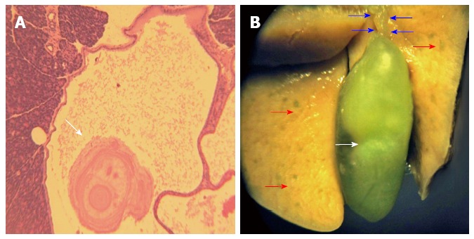
Figure 6.

Photomicrograph from pancreatic ductal calculi formation (A), as well as hepatic and gall bladder gross pathological structure (B) in dibutyltin dichloride-treated animals. A: Calculi formation: Photomicrograph illustrates histopathologic structure from a representative pancreas of a dibutyltin dichloride-treated animal to demonstrate ductal distension and calculi formation. Representative slide from n = 8 animals/group (magnification × 40); B: Photograph illustrates hepatic gross lesions (red arrows), expanded bile duct (blue arrows) and gall bladder (white arrow) from DBTC-treated animal. Representative from n = 8 animals/group.
