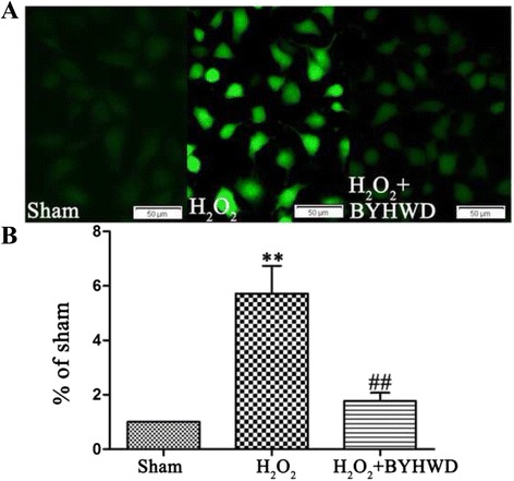
Fig. 6.

Effects of BYHWD on H2O2-induced intracellular ROS levels in HUVECs. a Representative images of intracellular ROS in HUVECs by fluorescence microscopy.b Quantitative analysis of intracellular ROS in HUVECs by spectrofluorophotometry. Values are shown as means ± SD (n = 10). ** P < 0.01, compared to sham group. ## P < 0.01, compared to H2O2 group (320 μmol/l)
