Fig. 4.

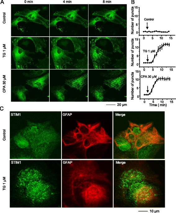
Depletion of calcium stores from endoplasmic reticulum induces STIM1 puncta formation in spinal astrocytes. a Live-cell confocal time-lapse images of STIM1-transfected astrocytes treated with 1 μM thapsigargin (TG) or 30 μM cyclopiazonic acid (CPA) at 0, 4, and 8 min. b Average number of puncta per 100 μm2 induced by respective treatments. Puncta were quantified as spots of high fluorescence intensity ranging from 0.4 to 2.0 μm in diameter size. c Confocal images of fixed spinal astrocytes containing endogenous STIM1 puncta after 8 min in the presence and absence of TG (1 μM)
