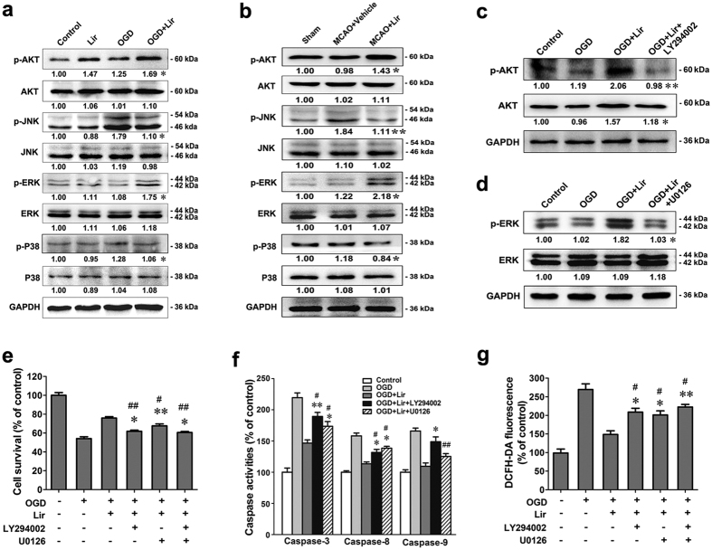
Figure 6. Liraglutide protects neurons by activating the PI3K/AKT and MAPK pathways.
(a) Western blot analysis of the expression of AKT, p-AKT, ERK, p-ERK, p38, p-p38, JNK and p-JNK in primary neurons (n = 3 experiments, *p < 0.05 vs. OGD group). n = 3 experiments, MCAO + vehicle group). (c) Expression of AKT and p-AKT in neurons after treatment with Lir and the PI3K inhibitor LY294002 in vitro (n = 3 experiments, **p < 0.01 vs. OGD + Lir group). (d) Expression of ERK and p-ERK after treatment with the Lir and ERK inhibitor U0126 in vitro (n = 3 experiments, *p < 0.05 vs. OGD + Lir group). (e) Cell viability was measured by the CCK-8 method following treatment with LY294002 and/or U0126. (f) The caspase activity of neurons treated with LY294002 and U0126 in vitro. (g) The intracellular ROS level measured by the DCFH-DA fluorescence intensity of neurons treated with LY294002 and/or U0126 after exposure to OGD for 60 min (n = 3 experiments, *p < 0.05 and **p < 0.01 vs. OGD + Lir group, #p < 0.05 and ##p < 0.01 vs. OGD group).

