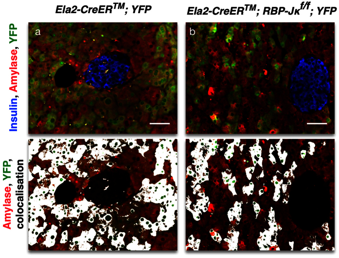
Figure 6. Lineage-tracing of RBP-Jκ deficient mature acinar cells.
Confocal imaging of immunostaining for Insulin (blue), Amylase (red) and YFP (green) on control (a) and Ela2-CreERTM; RBP-Jκf/f; YFP (in b) mice. The lower panels present the Amylase and YFP staining and overlays the colocalization in white. Scale = 50 μm.

