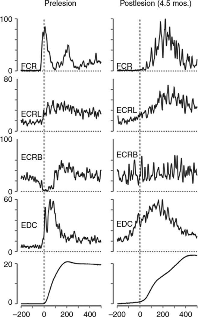
Figure 1.
Electromyographic (EMG) activity after an M1 lesion in an adult monkey. The monkey was trained to perform rapid step-tracking movements of the wrist to eight different target locations. EMG activity was full-wave rectified and averaged before (prelesion, left column) and after (postlesion, right column) the lesion. Averages were normalized, and the maximum EMG activity observed in one of eight wrist movement directions represents 100%. Note that, prelesion, the agonist (flexor carpis radialis [FCR]) burst began 40 ms before movement onset and lasted 100 ms. Activity in synergistics (extensor carpis radialis longus [ECRL] and extensor digitorum communis [EDC]) began 20 ms before movement onset. Activity in antagonistic (extensor carpis radialis brevis [ECRB]) muscle was strongly suppressed. Note that, postlesion, the agonist burst in the FCR was delayed. Synergistics (ECRL and EDC) showed early activity, and the suppression of antagonistic activity in ECRB was absent. Bottom traces showed displacement of the wrist (in degrees). Reproduced, with permission, from Hoffman DS, Strick PL. Effects of a primary motor cortex lesion on step-tracking movements of the wrist. J Neurophysiol. 1995;73: 891–895. Copyright © 1995 by The American Physiological Society.

