Figure 7.

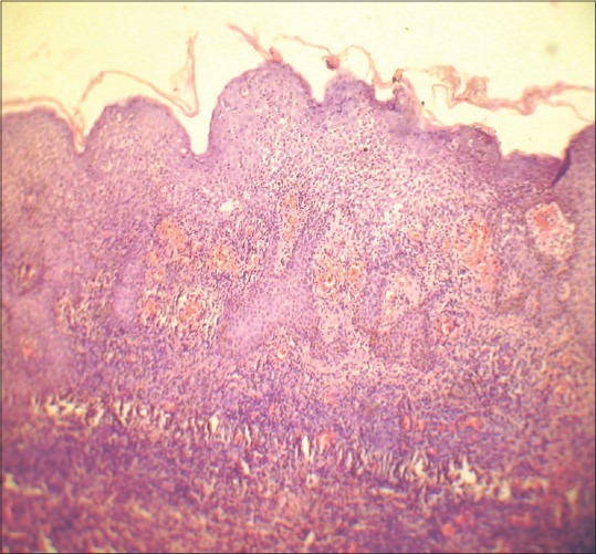
HPE (H and E stain, ×10) showing spongiosis, dense lymphocytic infiltration in the upper dermis around dilated blood vessels; some extravasated red blood cells may be seen

HPE (H and E stain, ×10) showing spongiosis, dense lymphocytic infiltration in the upper dermis around dilated blood vessels; some extravasated red blood cells may be seen