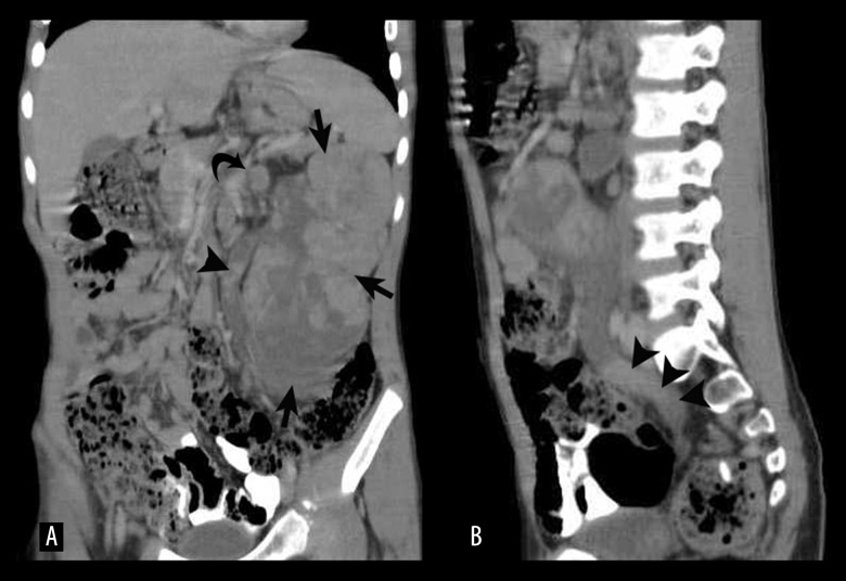
Figure 3.
(A) Coronal MPR image showing the tumour replacing the entire left kidney (arrows) along with a dilated ureter filled with multiple nodular deposits (arrowheads). Enlarged hilar nodes are also visualized (curved arrow). (B) Sagittal MPR image showing ureteric and renal pelvis dilatation with multiple tumour deposits (arrowheads).

