Figure 6.

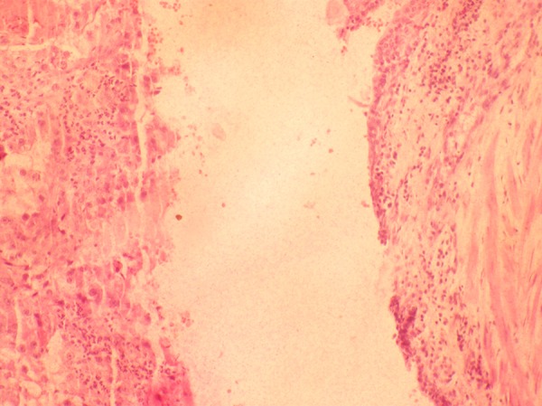
Haematoxylin and Eosin stain (10×) of a section from the resected margin of the ureter shows luminal dilatation, attenuated transitional cell lining epithelium and presence of tumour in the lumen.

Haematoxylin and Eosin stain (10×) of a section from the resected margin of the ureter shows luminal dilatation, attenuated transitional cell lining epithelium and presence of tumour in the lumen.