Figure 1.

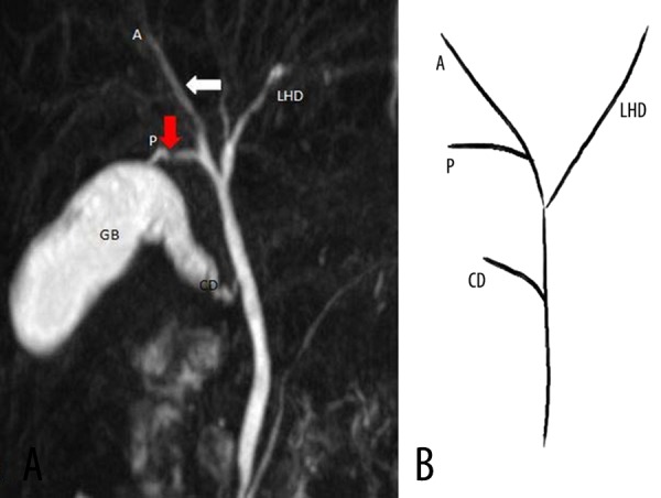
(A) Coronal 3D MR cholangiopancreatography, and (B) schematic diagram show RPSD (red arrow) joining the RASD (white arrow) from its lateral aspect. CD – Cystic duct, GB – gall bladder.

(A) Coronal 3D MR cholangiopancreatography, and (B) schematic diagram show RPSD (red arrow) joining the RASD (white arrow) from its lateral aspect. CD – Cystic duct, GB – gall bladder.