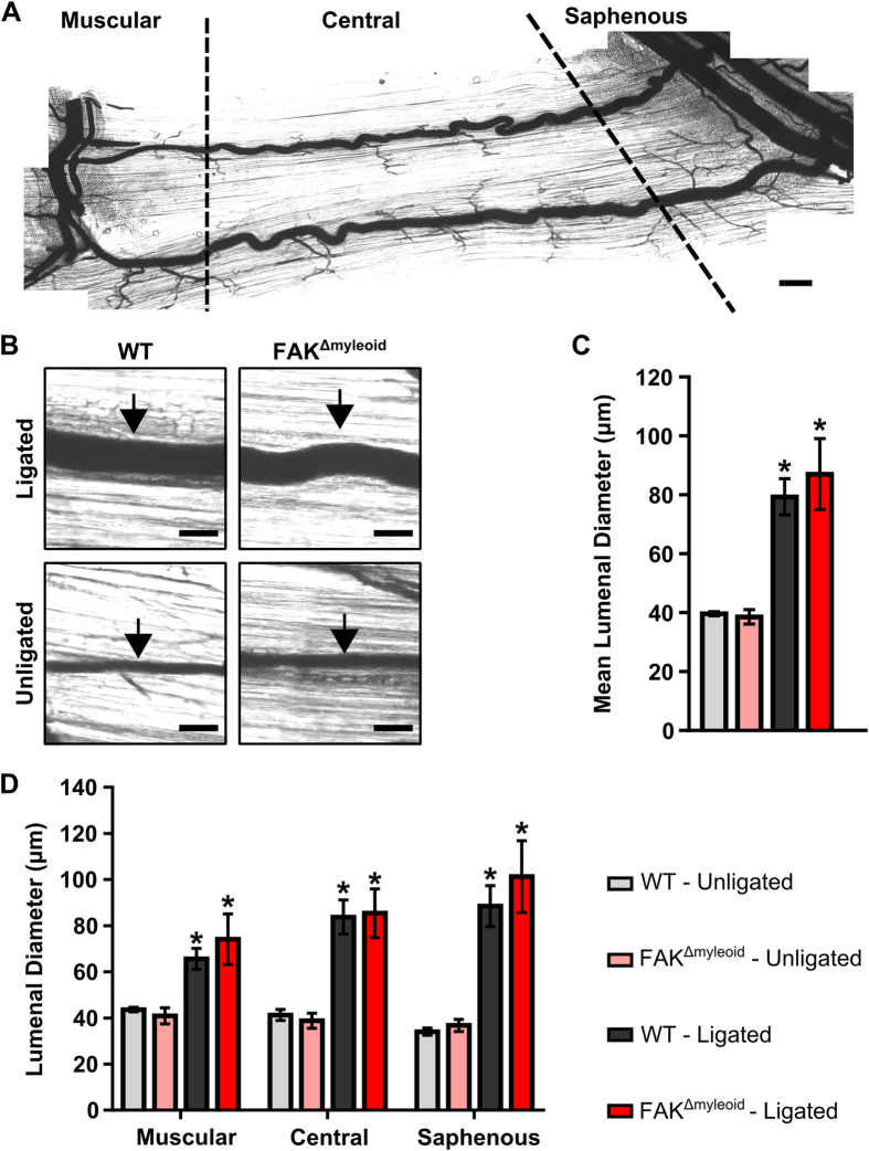
Figure 2. Arteriogenesis in the gracilis adductor muscle is independent of macrophage-specific FAK.
(A) Representative image of gracilis muscle whole mount preparation 28 days post-FAL indicating muscular, central, and saphenous regions of analysis separated by dotted lines. Scale bar = 500 μm (B) Representative images from central region of gracilis muscle whole mount preparations from the ligated and unligated limbs of FAKΔmyeloid and WT mice 28 days after FAL. Collateral arteries are identified by black arrows. Scale bar = 100 μm. (C) Bar graphs of mean lumenal collateral diameter averaged across all regions (muscular, central, and saphenous) and (D) in each region (muscular, central, and saphenous) between FAKΔmyeloid and WT mice. There were no statistical differences between FAKΔmyeloid and WT mice (n = 6). *p < 0.05 between ligated and unligated limbs within the given region.

