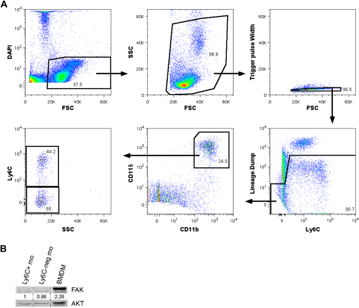
Figure 4. FAK is expressed at low levels in blood monocytes but is elevated in differentiated macrophages.
(A) Schematic of cell sorting gating procedure. Monocytes were identified as lineage negative (lineage defined as CD49b, CD3e, CD19, and Ly6G positive for NK cells, T cells, B cells, and neutrophils respectively), CD11b+, and CD115+. (B) Western blot of FAK expression in Ly6C+ monocytes (Ly6C+ mo), Ly6C-negative monocytes (Ly6Cneg mo), and bone marrow derived macrophages (BMDM). Ly6C+ monocytes and Ly6C-negative monocytes were isolated from wild-type mice via the cell sorting protocol described in (A). BMDMs were isolated from whole bone marrow and cultured in-vitro for 7 days in αMEM base media supplemented with 10% FBS, penicillin/streptomycin, and 10% CMG 14–12 conditioned media.

