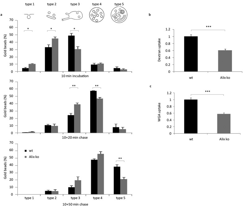
Figure 2. Endocytosis is delayed in Alix ko cells.
(a) EM quantification of BSA-gold particle endosomal trafficking. MEFs were incubated with BSA-gold for 10 min at 37 °C and fixed immediately (top panel), or washed and further incubated at 37 °C for 20 min (middle panel) or 50 min (bottom panel). 1000 gold particles were counted for each condition and were attributed to each compartment type: type 1: plasma membrane invaginations, type 2: endocytic vesicles, type 3: early endosomal compartments, type 4: multivesicular bodies (MVB), type 5: late endosomes/ lysosomes (*P < 0.05; **P < 0.01, two-tailed Student’s t-test). (b) MEFs were incubated 10 min with dextran-TRITC at 37 °C, washed and fixed. Mean fluorescent values per cell were estimated using ImageJ (Number of cells: wt, n = 123; Alix ko, n = 116 in 3 independent experiments ***P < 0.001, two-tailed Mann–Whitney U test). (c) MEFs were stained for 5 min with fluorescent WGA at 4 °C, washed and incubated 5 min at 37 °C. Mean fluorescent values per cell were estimated using ImageJ (Number of cells: wt, n = 114; Alix ko, n = 107 in 3 independent experiments ***P < 0.001, two-tailed Mann–Whitney U test).

