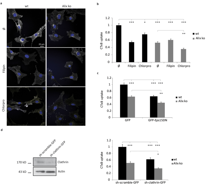
Figure 4. CIE but not CME of CTxB is impaired in Alix ko cells.
(a) Representative images of 5 min CTxB-TRITC uptake by wt and Alix ko MEFs. The reduced endocytosis of CTxB-TRITC seen in Alix ko cells is further decreased by chlorpromazine (chlorpro) but not by filipin. Ø: no treatment. Hoechst stained nuclei appear in blue. (b) Quantification of the experiment shown in (a). Mean fluorescence values per cell were calculated using ImageJ (Number of cells in 3 independent experiments: wt Ø, n = 107; wt filipin, n = 90; wt chlorpro, n = 72; Alix ko Ø, n = 145; Alix ko filipin, n = 100; Alix ko chlorpro, n = 95; *P < 0.05; **P < 0.01; ***P < 0.001, Dunn’s multiple comparison test). (c) Quantification of 5 min CTxB uptake by wt and Alix ko MEFs expressing GFP or GFP-Eps15DN. Mean fluorescence values per cell were calculated using ImageJ (Number of cells in 5 independent experiments: wt GFP, n = 147; wt GFP-Eps15DN, n = 93; Alix ko GFP, n = 147; Alix ko Eps15DN, n = 105; **P < 0.01; ***P < 0.001, Dunn’s multiple comparison test). (d) Quantification of CTxB uptake by wt and Alix ko MEFs expressing sh-clathrin-GFP. Wt and Alix ko MEF cells were transfected with an sh vector directed against clathrin (sh-clathrin-GFP) or a control vector (sh-scramble-GFP). 72 h later, cells were incubated with CTxB for 20 min at 4 °C, followed by 5 min at 37 °C and internalized CTxB was quantified. (Number of cells in 4 independent experiments: wt sh-scramble-GFP, n = 62; wt sh-clathrin-GFP, n = 68; Alix ko sh-scramble-GFP, n = 60; Alix ko sh-clathrin-GFP, n = 63; *P < 0.05; ***P < 0.001, Dunn’s multiple comparison test). Western blot analyses using anti-clathrin antibody demonstrate the efficacy of the sh-clathrin plasmid in downregulating clathrin expression in N2a cells 48 h after transfection.

