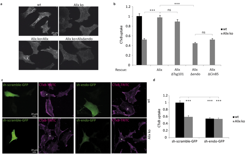
Figure 6. Alix and endophilin A2 mediate CIE of CTxB.
(a) Alix mediated CIE of CTxB requires binding to endophilins. CTxB-TRITC uptake by wt and Alix ko MEFs (upper panel) or by Alix ko cells transduced with viruses (lower panel) encoding for Alix (Alix ko + Alix), or for a mutant of Alix unable to bind to endophilin-A (Alix ko + AlixΔendo). Cells were incubated with CTxB during 20 min at 4 °C, followed by 5 min at 37 °C and acid stripping. (b) Quantification of CTxB uptake using the same type of experiments as shown in (a). Uptake of CTxB was quantified in wt or Alix ko cells or in Alix ko cells transduced with Alix or Alix deletion mutants lacking several amino-acids of the PRD corresponding to binding sites for Tsg101 (AlixΔTsg101: AlixΔP717–P720), endophilin-A (AlixΔendo: AlixΔP748–P761), or Cin85 (AlixΔCin85: AlixΔP739–R745). Mean fluorescence values per cell were estimated using ImageJ. (Number of cells in 4 independent experiments: wt, n = 104; Alix ko, n = 98; rescue Alix, n = 112; AlixΔTsg101, n = 117; AlixΔendo, n = 147; AlixΔCin85, n = 133. ***P < 0.001, Dunn’s multiple comparison test). (c) Endophilin-A2 and Alix act in a common pathway in CIE of CTxB. wt and Alix ko MEFs were transfected with plasmids containing sh-endophilin and coding for GFP (sh-endo-GFP) or a scrambled version of the same shRNA sequence (sh-scramble-GFP), and cultured for 72 h. Cells were then incubated with CTxB-TRITC (magenta) for 20 min at 4 °C, followed by 5 min at 37 °C. Surface bound toxin was removed by acid treatment prior to fixation and processing. Asterisks indicate the nuclei of transfected cells. (d) Quantification of the experiments shown in (c). CTxB mean fluorescence values per transfected cell were calculated using ImageJ software (Number of cells in 4 independent experiments: wt sh-scramble-GFP, n = 93; wt sh-endo-GFP, n = 70; Alix ko sh-scramble-GFP, n = 76; Alix ko sh-endo-GFP, n = 67. ***P < 0.001, one way ANOVA and Dunnett’s test).

