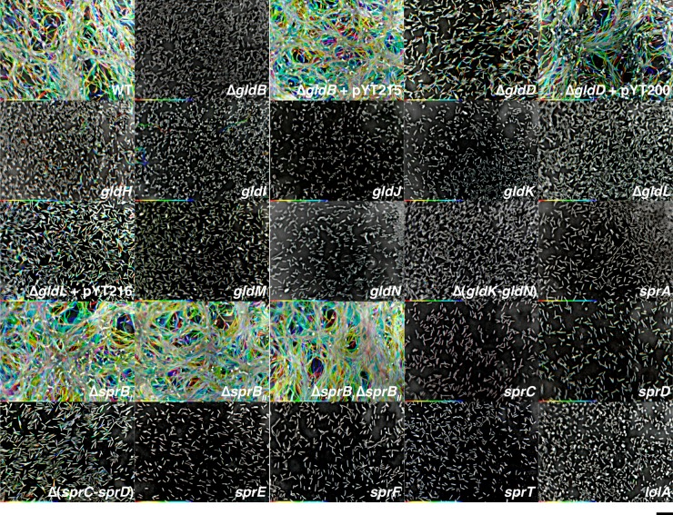
FIG 5.
Gliding of wild-type and mutant cells on agar. Cells were grown on Cytophaga agar at 15°C for 4 days, suspended in Cytophaga medium, and spotted on Cytophaga medium solidified with 1% agar on a glass slide. After drying, the cells were covered with an O2-permeable Teflon membrane that prevented dehydration and served as a coverslip. Cells were observed for motility at 20°C using an Olympus BH-2 phase-contrast microscope. A series of images were taken for 150 s using a Photometrics Cool-SNAPcf2 camera. Individual frames were colored from red (time zero) to yellow, green, cyan, and finally blue (150 s) and integrated into one image, resulting in rainbow traces of gliding cells. The rainbow traces correspond to the sequences shown in Movies S2, S4, and S6, and cells at time zero are shown in Fig. S4 in the supplemental material. Strains and plasmids are as listed in Fig. 3. White cells correspond to cells that exhibited little net movement. The scale bar indicates 10 μm and applies to all panels.

