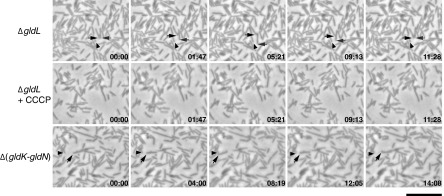
FIG 6.
Residual motility of C. algicola ΔgldL and Δ(gldK-gldN) mutants on agar. Cells in Cytophaga medium were spotted on Cytophaga medium solidified with 1% agar on a glass slide. After drying, the cells were covered with an O2-permeable Teflon membrane that prevented dehydration and served as a coverslip. Cells were observed for motility at 20°C using an Olympus BH-2 phase-contrast microscope. Individual frames are shown corresponding to C. algicola ΔgldL mutant CA150 (top row; taken from Movie S8 in the supplemental material), the ΔgldL mutant incubated with 20 μM CCCP to dissipate the proton gradient (middle row; taken from Movie S8), and Δ(gldK-gldN) mutant CA241 (bottom row; taken from Movie S9 in the supplemental material). Arrows indicate moving cells, and arrowheads indicate stationary reference points. Arrows and arrowheads were not added to the images of the ΔgldL mutant with CCCP because no cell movements were observed. The numbers indicate time in minutes:seconds. The scale bar indicates 10 μm and applies to all panels.

