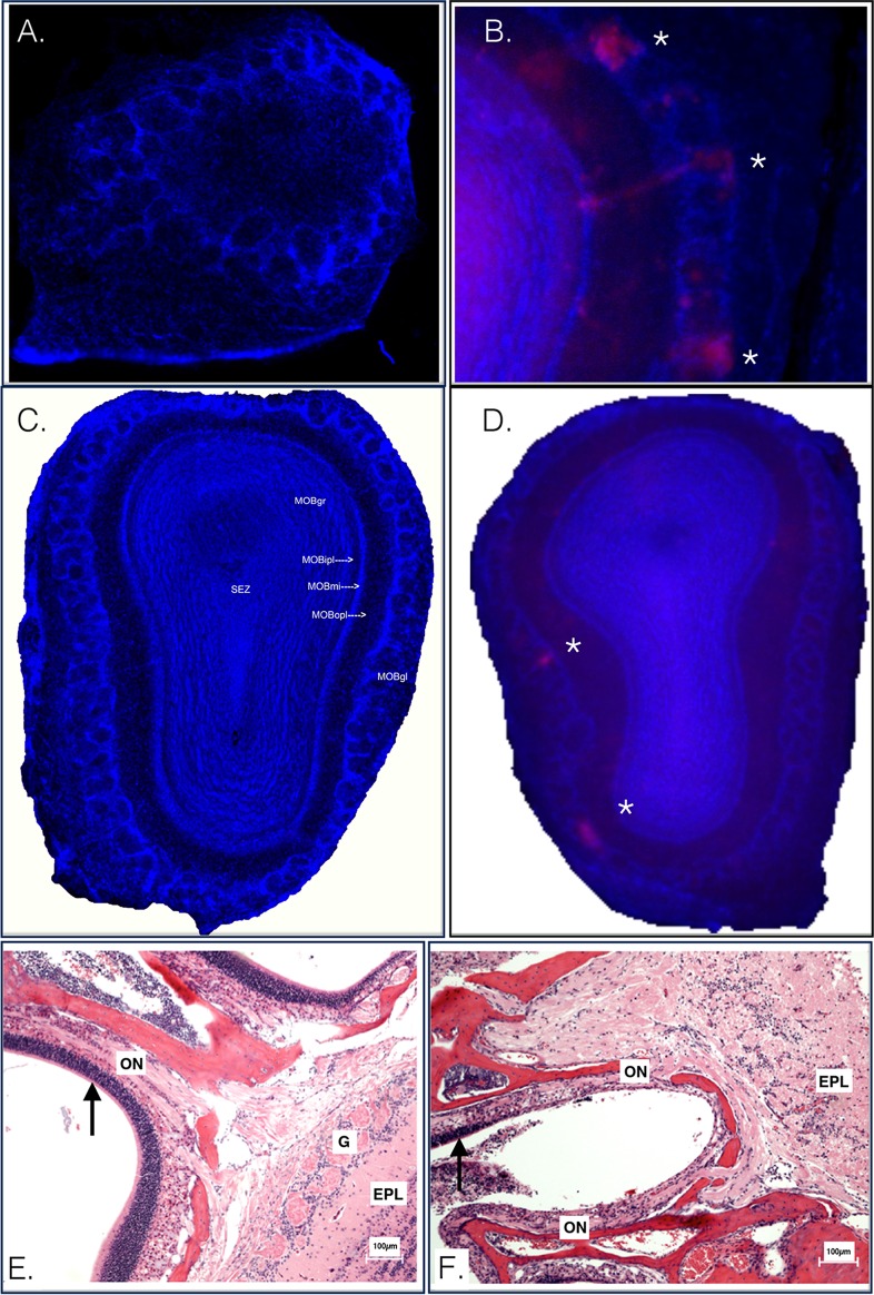
FIG 3.
A closer look at the olfactory bulb at early time points postinoculation. CD-1 mice were inoculated in the footpad with McRed virus and euthanized at 2 to 5 dpi. (A) CLARITY-treated olfactory bulb section imaged at a magnification of ×100, CD-1 mouse, footpad inoculation with McRed virus, 2 dpi. McRed virus is not observed in the olfactory bulbs of s.c.-inoculated animals. (B) Olfactory bulb section imaged at a total magnification of ×2, CD-1 mouse, intranasal inoculation with McRed virus, 2 dpi. Virus was readily detectable in the olfactory bulb glomerular layers of i.n.-inoculated mice. (C) CLARITY-treated olfactory bulb section imaged at a magnification of ×100, CD-1 mouse, footpad inoculation with McRed virus, 4 dpi. The montage image shows an olfactory bulb from a mouse with severe CNS infection, the same animal as shown in Fig. 6. Note that the olfactory bulb is pristine and devoid of viral expression. (D) Representative image for comparison showing McRed virus infection in the glomerular layer (asterisks) of the olfactory bulb following i.n. inoculation, 3 dpi. (E and F) Olfactory tract of CD-1 mice inoculated s.c. (E) or i.n. (F) with VEEV.3908 72 hpi. Arrows highlight the distinct difference in the condition of the epithelium of the nasal turbinates. EPL, external plexiform layer; G, glomerular layer; ON, olfactory nerve; MOBgr, main olfactory bulb granular layer; MOBipl, main olfactory bulb internal plaexiform layer; MOBmi, main olfactory bulb mitral cell layer; MOBopl, main olfactory bulb outer plexiform layer; MOBgl, main olfactory bulb glomerular layer.

