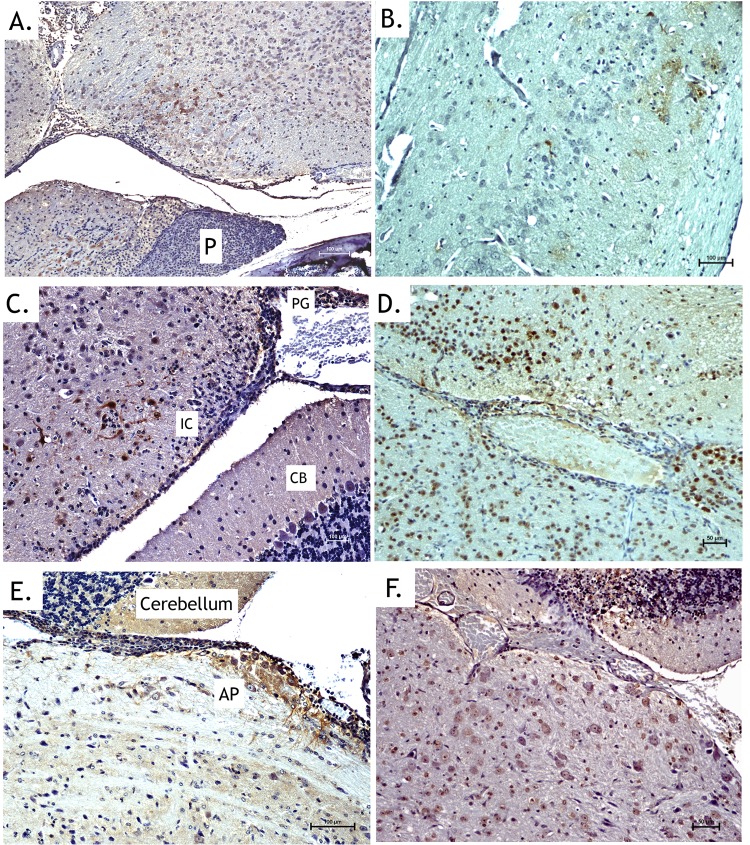
FIG 5.
Immunohistochemical staining supports in vivo imaging results by showing entry at CVOs. Bisected heads exhibiting increased luciferase activity were processed for histological analysis. Images presented are from animals euthanized at 3 to 4 days postinoculation. (A, C, and E) Anti-WEEV antigen immunohistochemical staining (brown) of the hypothalamic region with nearby pituitary gland (A), pineal body (C), and area postrema (E) following inoculation with McFire virus. (B, D, and F) Anti-luciferase immunohistochemical staining (brown) of the hypothalamus region (B), pineal body and inferior colliculus (D), and area postrema (F) following inoculation with VEEV-3908-fLUC. No staining occurred in the anterior pituitary (P). Staining was detected in the pineal gland (PG) and nearby neurons of the inferior colliculus but not in the cerebellum (CB). Strong staining of neurons and neuronal processes was observed in the area postrema. CTX, cortex.

