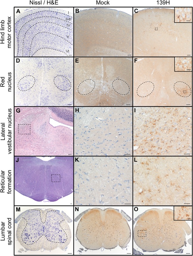
FIG 1.
Distribution of PrPSc in spinal cord, brain stem, and brain of hamsters infected in the sciatic nerve with the 139H agent. (A to C) Nissl staining (A) and PrPSc immunostaining (B and C) of the contralateral hind limb motor cortex from mock-infected (B) or 139H-infected (C) hamsters at 75 days postinfection. (D) Nissl staining of the mesencephalon containing the red nuclei (dashed outline). (E and F) PrPSc immunoreactivity was detected in the ventrolateral portion of the contralateral red nucleus at 75 days postinfection (F) but was not detected in mock-infected animals (E). (G) Hematoxylin and eosin (H&E) staining of the pons ipsilateral to the side of inoculation that contains the lateral vestibular nucleus. (H and I) At 100 days postinfection, PrPSc immunoreactivity is detected in the 139H-infected tissue (I) but is not detected in mock-infected animals (H). (J to L) H&E-stained section of the pons (J) containing the reticular formation contains PrPSc immunoreactivity in 139H-infected (L) but not in mock-infected (K) hamsters. (M) Nissl-stained section of lumbar spinal cord that contains ventral motor neurons (VMNs) whose axons are contained in the sciatic nerve. (N and O) PrPSc immunoreactivity of lumbar spinal cord from uninfected animals failed to detect PrPSc (N), while in 139H-infected hamsters at 50 days postinfection, PrPSc deposits were associated with VMNs ipsilateral to the side of inoculation (O). Scale bars, 200 μm in main panels and 100 μm in insets.

