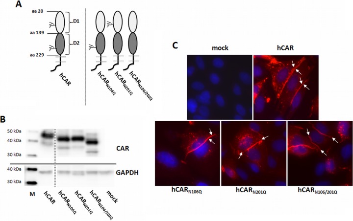
FIG 1.
Expression of hCAR glycosylation mutants in CHO-K1 cells. (A) Schematic illustration of wild-type hCAR and hCAR glycosylation mutants. (B) Protein expression in transiently transfected CHO-K1 cells analyzed by Western blotting with polyclonal anti-CAR rabbit antibody. Mutants lacking glycosylation of the D1 domain (hCARN106Q), the D2 domain (hCARN201Q), or both domains (hCARN106/201Q) show increased mobility compared to wild-type hCAR. CHO-K1 cells exposed to transfection reagent without plasmid DNA (mock) served as a negative control. All lanes are from a single exposure of a single immunoblot; the line indicates where irrelevant lanes were deleted. Lane M, MagicMark XP Western protein standard. (C) Localization of wild-type hCAR and hCAR mutants detected with anti-CAR rabbit antibody (red). DAPI was used for nuclear staining (blue). Both wild-type hCAR and hCAR glycosylation mutants were concentrated at cell-cell junctions (arrows). Magnification, ×400.

