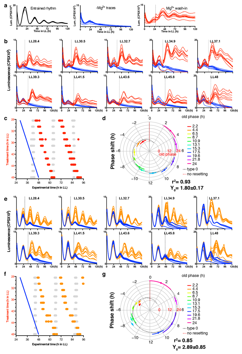
Extended Data Figure 8. Bioluminescence data of wedge experiment.
Peak expression phase of the clock protein CCA1 was analysed upon re-introduction of magnesium to cultures in low extracellular magnesium, to test whether the phase of cellular rhythms is dictated by the prior phase of entrainment or by this enforced transition from low to high [Mg2+]i. a, Bioluminescence traces showing that circadian rhythms in Ostreococcus are reversibly attenuated by depletion of extracellular Mg2+, and restored by Mg2+ wash-in. b,e. Bioluminescence traces from cells in low extracellular magnesium (b; 5 µM, e; 20 µM)with rhythms rescued by release into media containing normal physiological concentrations of magnesium at the indicated times (vertical dotted lines) in constant light (LL), compared to their respective controls where no magnesium was added in (blue traces). Data from 7-8 replicate wells are shown in each panel. c,f. Summary graphs where results from b,e are plotted in circadian wedge graphs: peak phases of CCA1-LUC rhythms in untreated control cells (grey dots) are compared with peak phase of rhythms reinstated by introduction of physiological magnesium following depletion to 5 µM (c, red dots) or 20 µM (f, orange dots), revealing that the phase of resulting rhythms is dictated solely by the phase of magnesium reintroduction (blue line). d,g. radial plots of phase shift (mean±SD, circumferential axis) depicted in panels c and f, versus phase prior to addition of Mg2+ to normal levels (old phase, radial axis and colour). The expected phase responses for type 0 resetting (black dotted line) and no resetting (red dotted line) are indicated. The goodness of fit (r2) and Y intercept (Y0) to the type 0 model are shown. Dose-dependent effects of intracellular magnesium on a critical clock parameter are confirmed by the observation that resetting is less strong when magnesium was reintroduced to cells adapted to intermediate levels of extracellular magnesium (e-g) compared to lowest extracellular magnesium (b-d).

