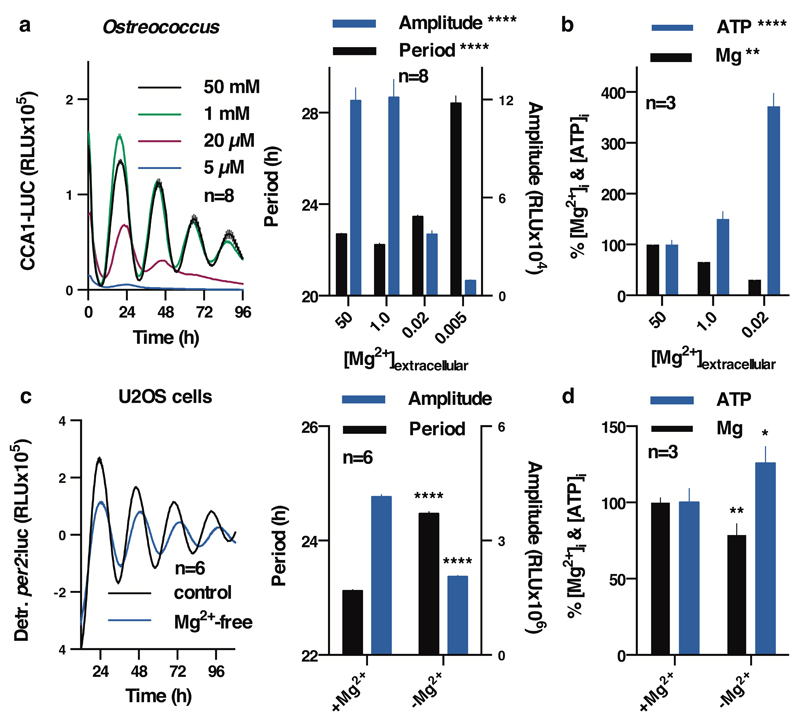
Figure 3. Reduced [Mg2+]i affects properties of cellular timekeeping and leads to an increase in ATP.
Bioluminescence traces showing reduced extracellular magnesium significantly affects amplitude and period length of circadian reporters in algal (a) and human cells (c). Low extracellular magnesium leads to decreased [Mg2+]i and increased [ATP]i in both cell types (b,d), measured after 4 days. All plots show mean±SEM, with replicate numbers (n) indicated, p-values (****p<0.0001, **p=0.01, *p=0.04) report significance by 1-way ANOVA (Ostreococcus) or t-test (U2OS).

