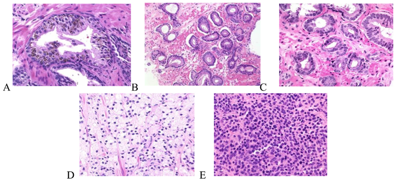
Figure 3.
Stained prostate tissue samples representing various stages of prostate cancer. Further details and description of changes in the tissue samples with progression of the disease is provided in the text. Reproduced with permission from http://www.webpathology.com.

