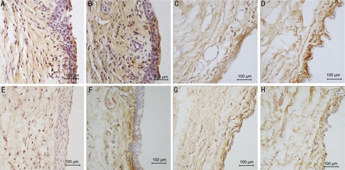
Figure 6. The immunohistochemical pictures show the expression of MMP-2 in conjunctiva and subconjunctival tissue of 10×40 magnification.
A: 3d group control eye, over-proliferative epithelial cells and fibroblasts in subconjunctiva, brown granules show the MMP-2 expression in all layers of tissues; B: 7d group control eye, brown staining in all layers; C: 21d group control eye, darker brown in subconjunctiva; D: 28d group control eye; E: 3d group olmesartan eye with milder brown in the same areas as the control; F: 7d group olmesartan eye, lighter brown mostly in the subconjunctival area right beneath the epithelial; G: 21d group olmesartan eye, lighter brown in all layers; H: 28d group olmesartan eye.

