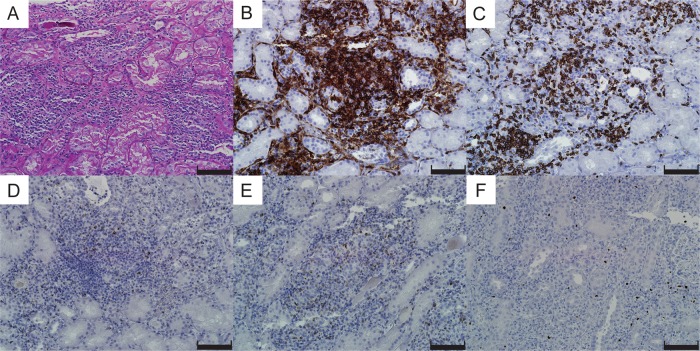
Fig. 2.
Light microscopy of the kidney biopsy: (A) Periodic acid–Schiff staining showed severe interstitial infiltration of inflammatory cells, mainly lymphocytes and neutrophils. (B–F) Immunohistochemistry revealed extensive T-cell infiltration of CD4+ (B) and CD8+ T cells (C). Ten to 20% of the infiltrates were positive for granzyme B (D) and perforin (E). Foxp3-positive regulatory T cells were also present (F). Scale bars, 100 μm.

