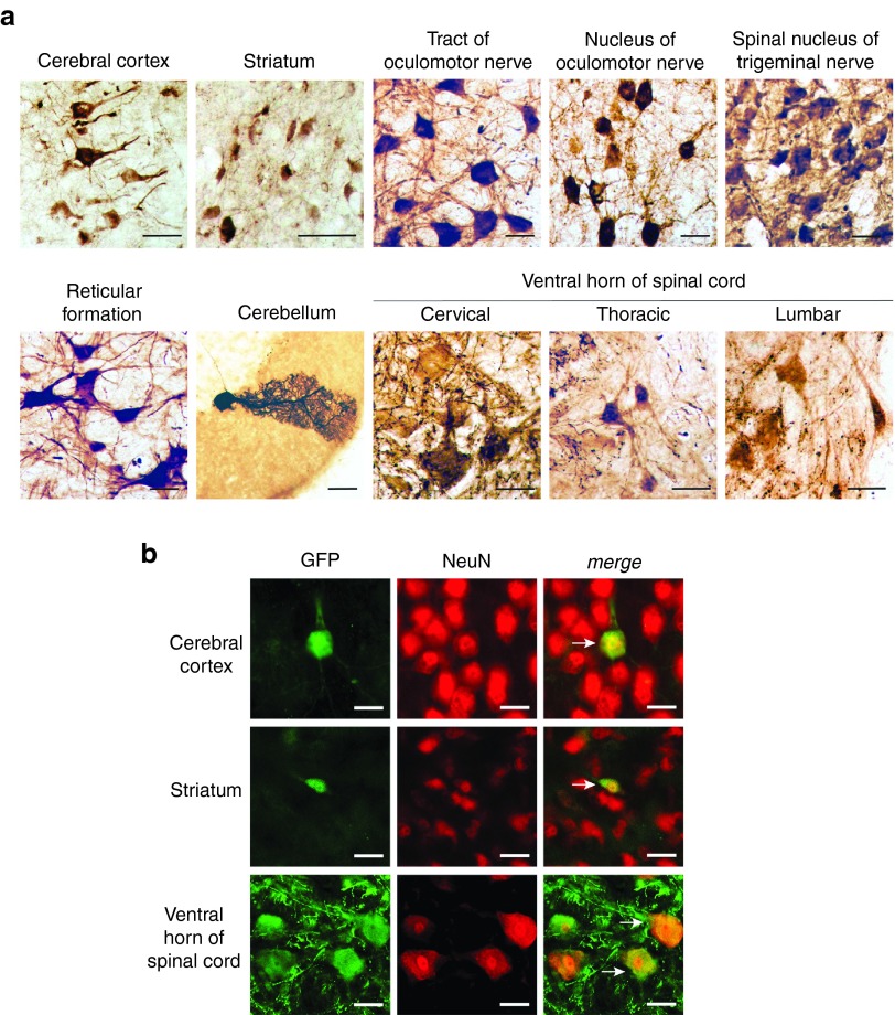
Figure 4.
Neuronal transduction in cat after systemic delivery of AAV-AS vectors. (a) Transduction of neurons in the cat brain after systemic delivery of AAV-AS vector (1.29 × 10¹³ vg). Representative images (left) show GFP-positive cells with neuronal morphology in various structures in the brain and spinal cord. Bar = 50 µm. (b) Double immunofluorescence staining for GFP and NeuN (right) confirm the neuronal identity of GFP-positive cells in brain and spinal cord. White arrows indicate examples of GFP-positive neurons. Bar = 50 µm.

