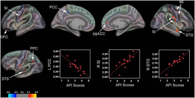
Fig 2. Brain Areas Showing Significant Associations between Cortical Thickness and Abdominal Pain Severity.
Brain areas showing changes in cortical thickness that were associated with abdominal pain severity rendered onto an inflated averaged brain for the left and right hemispheres (lateral and medial views) with corresponding scatter plots for selected clusters. Patients with greater abdominal pain complaints exhibited significant cortical thinning in the left posterior cingulate cortex (PCC) and cortical thickening in the left orbitofrontal cortex (OFC), bank of the superior temporal sulcus (STS), and posterior parietal cortex (PPC). The latter two clusters are displayed on an inflated surface of the left hemisphere that was tilted approximately 15° to highlight areas exhibiting cortical thickening. In the right hemisphere, abdominal pain severity was associated with cortical thickening in the superior temporal gyrus (STG), primary motor cortex (MI), and perigenual anterior cingulate cortex (pgACC), although the latter cluster did not pass the defined cluster-extent threshold. Patients also showed cortical thickening in the bilateral primary somatosensory cortices (SI) corresponding to the intra-abdominal or ‘viscerotopic’ area, with two clusters in the right hemisphere; an the inferiorly- and a superiorly-placed cluster. Scatter plots depicting correlations between cortical thickness values (mm) for significant clusters in the left (L) PCC, right (R) inferior SI intra-abdominal area, and R STG with respective abdominal pain severity scores.

