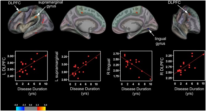
Fig 4. Cortical Thickness and Disease Duration in Pediatric IBS Patients.
Brain areas showing a significant relationship between cortical thickness and disease duration rendered onto an inflated averaged study-specific brain for the left (L) and right (R) hemispheres (lateral and medial views) with corresponding scatter plots for selected clusters. Top panel: Longer disease durations (yrs) were correlated with cortical thickening (warm colors) in the L and R DLPFC, and L supramarginal gyrus. In contrast, cortical thinning (cool colors) in the R lingual gyrus was correlated with disease durations in IBS patients. Bottom panel: Scatter plots illustrating positive and negative correlations between cortical thickness values (mm) for significant clusters and duration of IBS symptoms.

