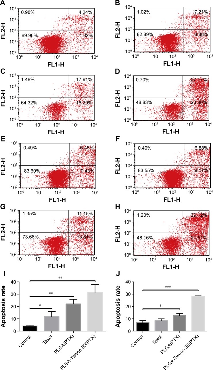
Figure 7.
PLGA-Tween 80 nanoparticles evidently induced cell apoptosis in both A549 and A549/T cell line.
Notes: Cell apoptosis analysis of A549 cells by flow cytometry using staining of Annexin V-FITC and PI treated with blank medium (A), Taxol (B), PTX-loaded PLGA NPs (C), and PTX-loaded PLGA-Tween 80 NPs (D) for 24 h. Cell apoptosis analysis of A549/T cells by flow cytometry using staining of Annexin V-FITC and PI treated with blank medium (E), Taxol (F), PTX-loaded PLGA NPs (G), and PTX-loaded PLGA-Tween 80 NPs (H) for 24 h. The percentage of double FITC and PI positive population of A549 cells (I) and A549/T cells (J) after treatment with Taxol, PTX-loaded PLGA NPs, and PTX-loaded PLGA-Tween 80 NPs for 24 h, respectively. *P<0.05; **P<0.01; ***P<0.001.
Abbreviations: PI, propidium iodide; PTX, paclitaxel; PLGA, poly(d,l-lactide-co-glycolide); NPs, nanoparticles; h, hours; V-FITC, fluorescein isothiocyanate.

