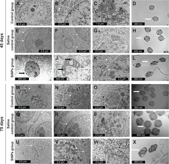
Figure 3.
The effects of SiNPs on the ultrastructure of testicular tissue in mice.
Notes: (A–D) The ultrastructure of testes in the control group on the 45th day after the first dose. (E–H) The ultrastructure of testes in the saline control group on the 45th day after the first dose. (I and J) SiNPs were observed in the SiNPs group on the 45th day after the first dose. (K and L) SiNPs led to swollen mitochondria and changed the shape of the cross-sections of the sperm tail in the SiNPs group on the 45th day after the first dose. (M–P) The ultrastructure of testes in the control group on the 75th day after the first dose. (Q–T) The ultrastructure of testes in the saline control group on the 75th day after the first dose. (U–X) The ultrastructure of testes in the SiNPs group on the 75th day after the first dose. The swollen mitochondria weren’t observed, and the shape of the cross-sections of the sperm tail were similar to that in the saline control group. The figure shows TEM images of testis. The thin black arrow represents the mitochondria, thin white arrow represents the cell membrane or nuclear membrane, wide white arrow represents the cross sections of the sperms, and wide black arrow represents the SiNPs. The data indicated that SiNPs could accumulate in the testis and damage the mitochondria and sperms.
Abbreviations: SiNPs, silica nanoparticles; TEM, transmission electron microscopy.

